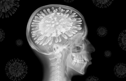
ATLANTA — So much focus on COVID-19 is placed on how the disease can impact a patient’s lungs and ability to breathe. Scientists say its effects on the brain shouldn’t be overlooked. New research performed by experts at Georgia State University and the Georgia Institute of Technology shows a decrease in the volume of gray matter in people with COVID-19 who develop a fever or get oxygen treatment.
According to their study, this reduction occurs specifically in the region between the frontal and temporal lobes of the brain. Reduced gray matter volume throughout this area was related to an increased level of impairment among those with COVID-19, even up to six months after being released from the hospital, according to the study.
Gray matter is essential for information processing. Abnormalities in gray matter may impair the efficiency by which neurons operate and interact. The findings suggest that the frontal lobe may contain gray matter involved in brain engagement in COVID-19, even outside of damage, like a stroke, caused by clinical symptoms of the illness.
Brain scans reveal differences between COVID patients, healthy people
Experts from the Center for Translational Research in Neuroimaging and Data Science (TReNDS) examined CT scans from 120 neurological patients. This group included 58 COVID-19 patients with severe symptoms and 62 healthy individuals who were all similar in age, gender, and medical history.
“Science has shown that the brain’s structure affects its function, and abnormal brain imaging has emerged as a major feature of COVID-19,” explains study co-author Kuaikuai Duan, a graduate research assistant at TReNDS and Ph.D. student in Georgia Tech’s School of Electrical and Computer Engineering, in a statement. “Previous studies have examined how the brain is affected by COVID-19 using a univariate approach, but ours is the first to use a multivariate, data-driven approach to link these changes to specific COVID-19 characteristics (for example fever and lack of oxygen) and outcome (disability level).”
Even after adjusting for cerebrovascular disorders, individuals with higher degrees of impairment had decreased gray matter volume in the upper, middle, and mid-frontal cortex at the time of discharge and up to six months later. The volume of gray matter in this area was similarly considerably decreased in oxygen therapy patients compared to non-oxygen therapy patients. When compared to individuals without fever, patients experiencing fever exhibited a substantial decrease in the volume of gray matter in the lower and mid-temporal region, as well as the fusiform gyrus.
Gray matters
The findings imply that COVID-19 may have an effect on the frontal-temporal system via fever or a shortage of oxygen.
Individuals with anxiety had less gray matter in the upper, middle, and mid-frontal cortex than those who were not anxious. This suggests that alterations of gray matter in the frontal lobe may be at the root of anxiety and depression seen in patients with COVID-19.
“Neurological complications are increasingly documented for patients with COVID-19,” explains Vince Calhoun, director of TReNDS and senior author of the study. “A reduction of gray matter has also been shown to be present in other mood disorders such as schizophrenia and is likely related to the way that gray matter influences neuron function.”
The results of the study show that alterations in the frontal-temporal system might be utilized as a biomarker to predict the outcome of COVID-19 or to assess therapeutic choices for the infection. Following that, the team wants to repeat the investigation using a larger sample with a variety of neurological scans and COVID-19 patient groups.
This study is published in Neurobiology of Stress.
