
CHICAGO, Ill. — Drinking during pregnancy is a combination doctors tell women to avoid at all costs. Now, a new study reveals that alcohol can alter the structure of an unborn baby’s brain.
A team with the Radiological Society of North America says drinking can lead to fetal alcohol syndrome (FAS), a range of developmental problems due to alcohol exposure while in the womb. The findings come from an analysis of 500 mothers-to-be in Austria undergoing MRI scans for clinical reasons.
“Fetal alcohol syndrome is a worldwide problem in countries where alcohol is freely available,” says lead author Professor Gregor Kasprian from the Medical University of Vienna in a media release. “It’s estimated that 9.8% of all pregnant women are consuming alcohol during pregnancy, and that number is likely underestimated.”
The team found significant changes in the brains of fetuses exposed to alcohol compared to healthy babies whose mothers avoided drinking.
“It appears that alcohol exposure during pregnancy puts the brain on a path of development that diverges from a normal trajectory,” Prof. Kasprian continues. “Fetal MRI is a very powerful tool to characterize brain development not only in genetic conditions, but also acquired conditions that result from exposure to toxic agents.”
How does alcohol affect a baby’s brain?
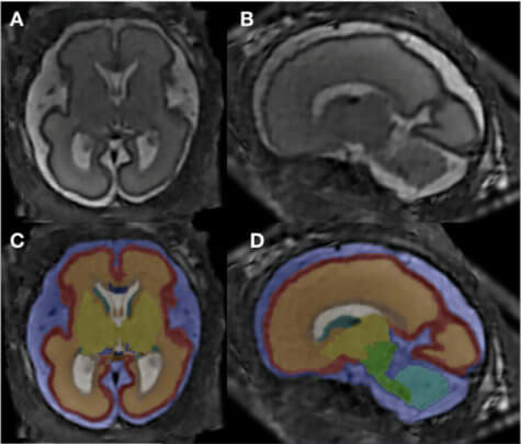
In particular, volume in two brain regions — the corpus collosum and periventricular zone — increased or decreased depending on alcohol consumption. The former is a large bundle of more than 200 million nerve fibers that communicate between the right and left sides of the brain. The latter sits at the base of the brain and is the organ’s neuron “factory.”
“One of the main hallmarks of our study is that we investigated so many smaller sub-compartments of the brain,” explains co-author Dr. Marlene Stuempflen.
FAS is the most severe form of a group of conditions doctors call fetal alcohol spectrum disorders.
Babies may have specific physical features such as small eyes, a thin upper lip, and a smooth area under the nose. Other problems include mental deficits, malformations of bones and major organs, inhibited growth, and central nervous system illnesses.
Children with FAS may also suffer from poor motor skills, higher mortality rates, and difficulties with learning, memory, social interaction, and attention span. One in 70 pregnancies with alcohol exposure results in FAS, according to Prof. Kasprian.
Mapping out the damage alcohol does to the brain
In the first study of its kind, 51 participants admitted consuming alcohol during their pregnancy. They took part in a surveillance project called PRAMS (Pregnancy Risk Assessment Monitoring System) and completed another questionnaire called T-ACE that screens for risky drinking behaviors.
“We provided a safe environment where women could feel comfortable honestly answering the questions,” Prof. Kasprian says.
The researchers combined statistical analysis with super-resolution imaging that created one dataset to re-construct each fetal brain. They completed 12 different brain structures, computing total and segment volumes of specific compartments.
“This is the first time that a prenatal imaging study has been able to quantify these early alcohol-associated changes,” adds Dr. Stuempflen.
No safe amount while pregnant
The corpus collosum is the main connection between the brain’s left and right sides. Dr. Stuempflen notes that it’s fitting this very central structure is affected by drinking since the clinical symptoms of FAS disorders are highly diverse. They cannot be pinpointed to one specific substructure of the brain.
“The changes found in the periventricular zone, where all neurons are born, also reflect a global effect on brain development and function,” Dr. Stuempflen reports.
Identifying a thicker corpus collosum in the alcohol-positive fetuses was surprising, as it’s thinner in infants with FAS disorders.
“There are many postnatal studies on infants exposed to alcohol,” Dr. Kasprian notes. “We wanted to see how early it’s possible to find changes in the fetal brain as a result of alcohol exposure.”
Study authors eliminated some of the fetal MRIs due to structural brain anomalies and poor image quality. The final group consisted of 26 scans from 24 alcohol-positive fetuses and a control group of 52 gender and age-matched healthy peers. At the time of imaging, the fetuses ranged in age from 20 to 37 weeks.
Both the CDC and previous studies have concluded that there is no safe amount of alcohol a woman can have while pregnant or even while trying to get pregnant. Alcohol passes from the mother’s blood to their baby through the umbilical cord. Along with FAS, the CDC warns that drinking while pregnant can also lead to miscarriage or stillbirth.
The researchers presented their findings at the annual meeting of the Radiological Society of North America.
South West News Service writer Mark Waghorn contributed to this report.