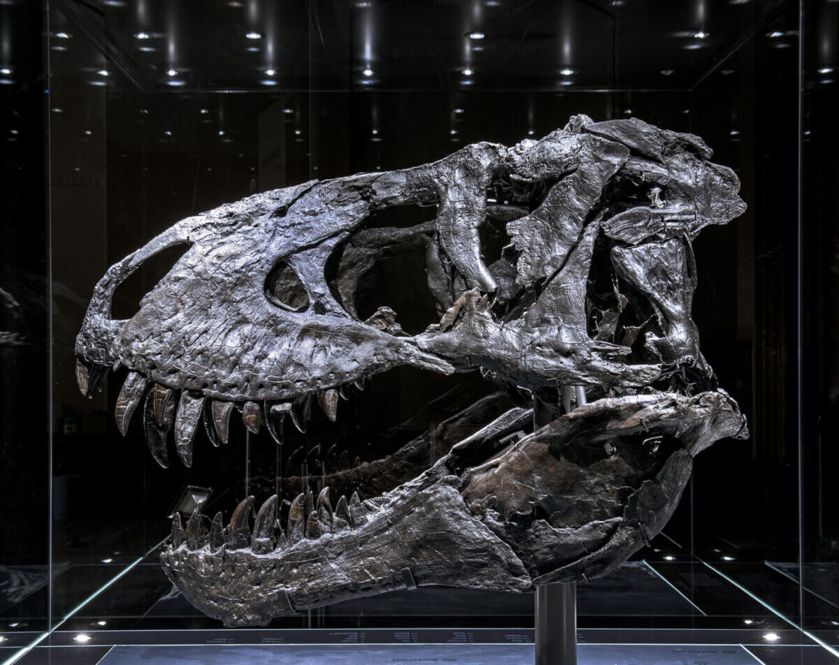
The “Tristan Otto” Tyrannosaurus rex skull that was examined by researchers.
CHICAGO, Ill. — A Tyrannosaurus rex that roamed the Earth 68 million years ago appears to have had a bone disease that would have caused severe tooth pain, a new study reveals. A team from Germany says the severe infection, called tumefactive osteomyelitis, originated in the marrow of the dinosaur’s left jaw.
It would likely have given the beast, which scientists nicknamed “Tristan Otto,” an agonizing toothache — turning it into a particularly bad-tempered predator. The creature’s fossilized remains are almost completely intact, making it one of the most well-preserved specimens ever discovered.
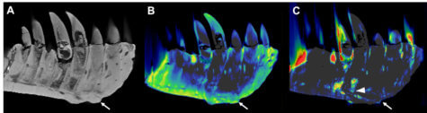
Now, scans have identified one of the earliest known cases of a painful condition that regularly affects humans. Lead author Dr. Charlie Hamm, a radiologist at Charité University Hospital in Berlin, says a review of CT imaging of this fossil revealed thickening along the left dentary and a mass on the jaw surface that extended to the root of one of Otto’s teeth.
The German team used a non-invasive technique called DECT (dual-energy computed tomography) to make this discovery. The scans detected a significant accumulation of the element fluorine, a finding linked to brittle bones.
Dr. Hamm adds the mass and fluorine buildup supports the diagnosis of tumefactive osteomyelitis, an infection of the bone.
This dinosaur was bigger than a bus
Paleontologists discovered the T-rex in Montana in 2010. It is one of only a few T-Rex skulls with a complete set of its 60 lethal dagger-like teeth. At 13 feet tall, 40 feet long, and weighing about eight tons, Otto is bigger than a double decker bus.
Only about 50 T-Rex fossils have been unearthed since the first was discovered in 1902. Not one has been found 100 percent intact. With 170 original bones out of the roughly 300 parts in the skeleton, Tristan Otto is among the best specimens scientists have to learn from.
The researchers described T-Rex as a “familiar subject of today’s popular culture.” Researchers believe it was one of the largest predators to ever walk the Earth. The creature’s notoriety has led to it being dubbed the “king of the dinosaurs.”
Massive jaws unleashed a bite so strong they turned armor plated animals into a packed lunch, chomping down with a force of over six tons. However, the new findings suggest that this severe toothache would have made Tristan Otto an even angrier foe as it hunted around the present-day western United States.
DECT preserves fossils better than other scanning methods
The imaging method has important implications in paleontology as an alternative to assessment methods that damage fossil samples. Dr. Hamm explained DECT deploys X-rays at two different energy levels to provide information about tissue composition and disease processes.
“We hypothesized that DECT could potentially allow for quantitative noninvasive element-based material decomposition and thereby help paleontologists in characterizing unique fossils,” Dr. Hamm says in a media release.
The researchers were able to overcome the difficulties of scanning a large portion of Tristan Otto’s lower jaw. The piece’s compactness was particularly challenging as imaging quality suffers when looking at very dense objects.
“We needed to adjust the CT scanner’s tube current and voltage in order to minimize artifacts and improve image quality,” Dr. Hamm adds. “While this is a proof-of-concept study, noninvasive DECT imaging that provides structural and molecular information on unique fossil objects has the potential to address an unmet need in paleontology, avoiding defragmentation or destruction.”
Tristan Otto was on display at the Natural History Museum in Berlin for four years.
“The DECT approach has promise in other paleontological applications, such as age determination and differentiation of actual bone from replicas,” says the museum’s vertebrate paleontologist Dr. Oliver Hampe. “The experimental design, including the use of a clinical CT scanner, will allow for broad applications.”
Taking another look at ‘Sue’
Dr. Hamm and his colleagues also collaborated with U.S. paleontologists from to perform a CT analysis of “Sue,” the world-famous T-rex housed in the Field Museum in Chicago.
“With every project, our collaborative network grew and evolved into a truly multidisciplinary group of experts in geology, mineralogy, paleontology and radiology, emphasizing the potential and relevance of the results to different scientific fields,” Dr. Hamm concludes.
The team presented their findings at the annual meeting of the Radiological Society of North America (RSNA).
South West News Service writer Mark Waghorn contributed to this report.


